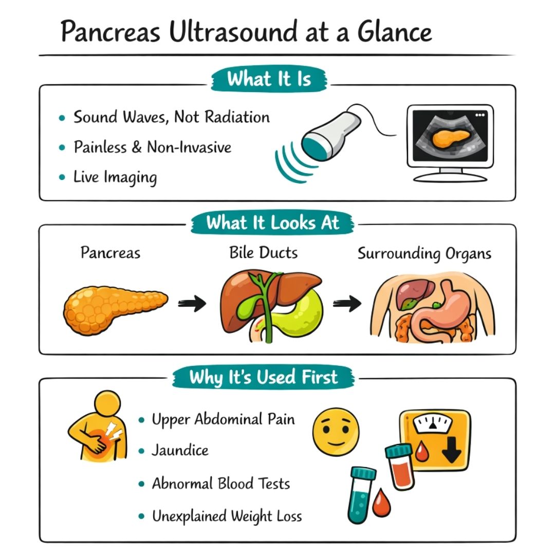
A pancreas ultrasound is a completely safe and painless imaging scan. It uses high-frequency sound waves, much like sonar, to create a live picture of your pancreas, giving doctors a first look at what’s going on without any radiation. It’s often the go-to starting point when investigating symptoms like unexplained pain in your upper abdomen or jaundice.
Your First Look Inside the Abdomen
If your doctor has suggested a pancreas ultrasound, it's completely normal to feel a bit anxious or have a lot of questions. This guide is here to walk you through exactly what happens, from start to finish, so you know just what to expect.
Think of your pancreas as a vital but shy organ, tucked away deep in your abdomen right behind your stomach. This hidden spot can make it tricky to examine. An ultrasound offers a clever, non-invasive way for your medical team to see the pancreas and the tissues around it clearly.
What Is This Scan Looking For?
So, why has your doctor ordered this particular scan? A pancreas ultrasound is a fantastic diagnostic tool for getting to the bottom of various symptoms and conditions. Its main job is to give your doctor a clear visual that helps them piece together what’s happening inside your body.
Some of the most common reasons include:
Investigating Abdominal Pain: If you're dealing with persistent or sharp pain in your upper belly, an ultrasound can help spot signs of inflammation or other problems.
Unexplained Weight Loss: Losing weight without intending to can be a red flag. This scan helps doctors check for underlying issues within your digestive system.
Symptoms of Jaundice: A yellow tinge to your skin or eyes often points to a blockage in the bile ducts, which are located near the pancreas. An ultrasound is great at visualising these ducts.
Abnormal Blood Test Results: If blood tests show unusual levels of certain enzymes, it might suggest the pancreas is under stress. A scan provides a much-needed closer look.
A pancreas ultrasound often acts as the first-line investigation. It’s like sending in a scout to gather initial intelligence, which then helps guide any further diagnostic steps and shapes your treatment plan.
Pancreatic ultrasounds are a cornerstone of the initial assessment for pancreatic diseases globally. To give you an idea of how common they are, abdominal and pelvic ultrasounds (which include imaging the pancreas) account for hundreds of thousands of procedures each month in many countries. This really highlights how much healthcare systems rely on this safe and effective method.
Getting to the root cause of symptoms is a crucial first step. While this scan focuses on the pancreas, it’s worth remembering that symptoms like unexplained weight loss can be linked to other conditions. You can learn more by reading our guide on colon cancer symptoms. Think of this ultrasound as one of the most important first steps toward getting the answers you and your doctor need.
Why Your Doctor Recommended This Scan
Hearing you need any kind of medical scan can be unsettling, but understanding exactly why your doctor has recommended it can make a huge difference. If a pancreas ultrasound is on the cards, it’s because your doctor is carefully piecing together clues from your symptoms to get a clearer picture of your health.
Think of it as the first, and often most straightforward, step in investigating what’s going on in your upper abdomen. It’s a completely safe way to get a direct look at your pancreas and the organs around it, helping to either confirm or rule out a number of potential issues. It's the initial look that guides every decision that comes next.
Connecting Symptoms to a Diagnosis
Your body is pretty good at sending signals when something isn’t quite right. A pancreas ultrasound helps your medical team translate those signals into real, actionable information. Your doctor likely suggested this scan to look into specific symptoms that point towards the pancreas or nearby organs.
Here are some of the most common reasons a pancreas ultrasound is ordered:
Persistent Upper Abdominal Pain: A nagging ache, tenderness, or even a sharp pain in your upper belly that sometimes spreads to your back is a classic sign something’s up with the pancreas. The scan can show inflammation or other changes causing this discomfort.
Jaundice: If your skin or the whites of your eyes have started to look yellow, it could mean a blockage in your bile ducts. Since these ducts run right through the pancreas, an ultrasound is a great way to check for obstructions like gallstones or a mass.
Unexplained Nausea or Vomiting: When you’re dealing with digestive problems like these without an obvious cause, looking at the pancreas can help figure out if it's the root of the problem.
Sudden, Unintended Weight Loss: This is a big one and always needs investigating. An ultrasound can assess the pancreas for any abnormalities that might be interfering with how your body digests food and absorbs nutrients.
Identifying Pancreatitis and Other Conditions
One of the most common reasons for a pancreas ultrasound is to check for pancreatitis, inflammation of the pancreas. This condition can be acute (coming on suddenly and severely) or chronic (lasting a long time). The scan allows doctors to see if the organ looks swollen or if there’s fluid around it, which are classic signs of inflammation.
In fact, cases of acute pancreatitis are a significant health concern, with an incidence rate between 15 to 42 cases per 100,000 people each year in some Western countries. Studies have shown just how essential ultrasound is for spotting gallstones and blockages in the bile ducts, which are major triggers for pancreatitis.
An ultrasound is a crucial first piece of the diagnostic puzzle. It gives your medical team foundational images that can immediately spot common problems like inflammation or cysts, helping them quickly decide on the best next steps for your care.
Beyond just inflammation, the scan is brilliant at identifying other structural issues:
Pancreatic Cysts: These are little fluid-filled sacs that can form on or inside the pancreas. Most are harmless, but some have the potential to cause trouble down the line, so getting a look at them is important.
Potential Tumours or Masses: The ultrasound can also spot abnormal growths. While it can't diagnose cancer on its own, finding a mass is a critical first step that will lead to more detailed imaging, like a CT or MRI scan. It's worth remembering that abdominal symptoms can sometimes be linked to other conditions. For a wider perspective, you might find our guide on primary peritoneal cancer helpful.
If you want to better understand why your doctor recommended this scan, or just need general health advice about your pancreas, you could chat with an online primary care provider. They can help make sense of your symptoms and walk you through the diagnostic process. Ultimately, this scan is a vital tool for narrowing down the possibilities and getting you on the right path to an accurate diagnosis and treatment.
How to Prepare for Your Appointment
Knowing what to do before your pancreas ultrasound can make a real difference, helping the process go smoothly and ensuring the sonographer gets the clearest possible images. The good news is that the prep is very straightforward.
The single most important thing you need to do is fast before your appointment. This means no eating or drinking for about 6 to 8 hours beforehand. You can usually take essential medications with a small sip of water, but it's always wise to double-check this with your doctor or the imaging centre.
Why Fasting is So Important
This isn't just a casual suggestion; it's crucial for getting a good scan. When you eat or drink, your digestive system kicks into gear, and this activity produces gas in your stomach and intestines. For an ultrasound, gas is the enemy.
Think of it like trying to see through a foggy window. The view is obscured and blurry. Gas in your abdomen does the same thing to the sound waves, blocking them and making it incredibly difficult for the sonographer to get a clear, detailed picture of your pancreas. By fasting, you give them a clear, unobstructed path.
What to Wear and What to Bring
On the day of the scan, your comfort is a priority. I always recommend wearing loose-fitting, two-piece clothing, like a top with trousers or a skirt. You'll likely be asked to lift your top and lower your waistband to expose your abdomen, so easy-to-adjust clothing just makes things simpler for everyone.
When you head to your appointment, make sure you have a few things with you:
Your appointment letter and any other hospital information.
A list of any medications you are currently taking. This gives the medical team a complete picture of your health.
Details of any known allergies, which is standard practice for any medical procedure.
Following the fasting instructions is the most critical part of your preparation. An empty stomach provides the best possible conditions for the ultrasound, helping your medical team capture high-quality images for an accurate assessment.
On the Day of Your Scan
Once you arrive at the clinic or hospital, the process itself is quite simple. A sonographer, who is a specialist trained in ultrasound imaging, will come and greet you. They’ll explain what's about to happen and get you settled.
They will ask you to lie down on an examination table, usually on your back, and make sure you're comfortable. Next, they'll apply a clear, water-based gel to your abdomen. It might feel a bit cool at first, but it warms up quickly from your body heat. The whole point of the gel is to get rid of any tiny air pockets between your skin and the ultrasound probe (called a transducer), which helps the sound waves travel properly into your body.
The sonographer will then gently press the transducer against your skin, moving it around your upper abdomen to find the best views. You might feel some light pressure, but it shouldn't be painful at all. They may ask you to take a deep breath and hold it for a few seconds. This simple action can help move other organs out of the way, giving them an even better look at your pancreas.
The entire scan usually takes around 30 minutes from start to finish. All you have to do is lie back and relax while the sonographer does their work.
Making Sense of Your Ultrasound Results
Once your pancreas ultrasound is finished, the job isn't quite done. The images are sent over to a radiologist – a doctor who is an expert in reading and interpreting medical scans. They’ll carefully examine every picture to build a detailed report. Let’s break down what they're looking for, turning that medical jargon into something much clearer.
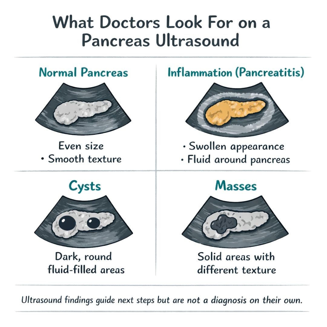
The first thing the radiologist does is get a general sense of your pancreas. A healthy one usually looks pretty uniform on the screen, with a consistent size, a clear shape, and a smooth texture. They will measure it and check its overall structure to see if everything looks as it should.
This image gives a simple overview of the key steps you’ll take on the day of your scan.
As you can see, the preparation, like fasting, is a vital first step. It clears the way for the best possible images during the scan itself.
What Different Findings Might Look Like
The radiologist’s real work is spotting anything that looks out of the ordinary. Different health conditions create unique visual signatures on an ultrasound, and understanding these can help you make sense of the report you might receive.
Here are a few common findings and what they could mean:
Inflammation (Pancreatitis): If your pancreas is inflamed, it often looks swollen and bigger than normal. Its texture might appear uneven or patchy, and sometimes the radiologist can see fluid collecting around it.
Cysts: A pancreatic cyst typically shows up as a dark, well-defined circle. Because it’s filled with fluid, it looks distinct from the solid tissue around it. The report will note its size and location, as most are harmless but some need to be watched over time.
Masses or Tumours: A solid growth will appear as a specific area that looks different from the surrounding pancreas—often darker and with a different texture. The radiologist will describe its size, shape, and whether it has clear, defined edges.
It’s crucial to remember that an ultrasound is an excellent starting point, but it's just one piece of the puzzle. Finding a mass, for example, doesn't automatically mean it's cancer. To understand more, our article on whether an ultrasound can show cancer offers some valuable extra context.
A key thing to remember is that an ultrasound report describes what the radiologist sees. It doesn't diagnose. That final interpretation comes from your doctor, who will connect the scan results with everything else they know about your health.
The Bigger Diagnostic Picture
Think of your ultrasound report as a set of detailed observations. The radiologist provides the facts, but it’s your own doctor who puts those facts into the context of you. They’ll look at everything together to build a complete picture.
This includes:
Your Symptoms: How do the findings on the scan match up with the abdominal pain, jaundice, or other issues you’ve been feeling?
Physical Examination: What did your doctor observe during your appointment?
Blood Test Results: Do your pancreatic enzyme levels or other blood markers support what the ultrasound is showing?
For example, if the ultrasound reveals a swollen pancreas and your blood tests show sky-high enzyme levels, a diagnosis of pancreatitis becomes very likely. On the other hand, if a suspicious mass is found, your doctor will almost certainly order more advanced scans to get a clearer, more definitive answer.
Next Steps After Your Results
Once your doctor has reviewed all this information, they’ll sit down with you to discuss what it all means. This conversation is the most important part of the process – it's your chance to ask questions and get a clear understanding of what’s next.
Depending on what was found, the next steps could be anything from dietary changes for mild pancreatitis to further imaging like a CT or MRI scan to get a better look at a cyst or mass. The ultrasound provides that vital first glimpse, pointing your medical team in the right direction for an accurate diagnosis and the most effective treatment plan.
The Role of Ultrasound in Pancreatic Cancer Care
Hearing the words "pancreatic cancer" is always a serious and worrying moment. If your symptoms or other test results have raised a concern, a pancreas ultrasound is very often the first imaging test your doctor will suggest. Think of it as the crucial starting point for a careful and thorough investigation, providing the initial look that will guide every step that follows.
The scan’s main job is to gather fundamental information quickly and safely. It’s the first piece of the diagnostic puzzle, giving your medical team a window into what’s happening inside your abdomen without using any radiation or invasive procedures. This initial view can provide immediate clarity and direction for what needs to happen next.
What Sonographers Look For
During the scan, the sonographer and radiologist are hunting for specific visual clues that might hint at a tumour. Their trained eyes are focused on identifying any abnormalities in the pancreas’s size, shape, or texture.
They are specifically on the lookout for:
A Distinct Mass: A tumour often shows up as a defined area that looks different from the healthy pancreatic tissue around it. On the screen, it might appear darker (a term we call hypoechoic).
Blocked Bile or Pancreatic Ducts: The pancreas is filled with tiny tubes, or ducts, that carry digestive juices. A growing tumour can press on these ducts, causing them to widen from the blockage—a tell-tale sign that an ultrasound can easily spot.
Changes in the Pancreas Shape: A growth can distort the normal, smooth outline of the pancreas. This is another subtle change that a specialist will be looking for.
Pancreatic cancer is one of the leading causes of cancer-related deaths worldwide. Tragically, because the early symptoms are often so subtle, many patients are diagnosed at a late stage, which has a major impact on their prognosis. The National Pancreatic Cancer Audit scoping report offers more insight into the importance of early detection and the current state of care.
Understanding the Limitations of Ultrasound
While an abdominal ultrasound is an invaluable first-line tool, it's important to be realistic about its limitations, particularly when we're talking about cancer. The pancreas is tucked away deep inside the abdomen, hiding behind the stomach and intestines. This positioning can sometimes make getting a crystal-clear view of the entire organ a real challenge.
Gas in the intestines can also get in the way, acting like a shield that blocks the sound waves. Because of these factors, a standard ultrasound might not be able to spot very small tumours, especially any located in the tail of the pancreas, which is the hardest section to see.
Think of a standard pancreas ultrasound as an initial survey of the landscape. It's excellent at spotting the larger, more obvious features, but it might miss smaller details hidden from view. It gives us the essential first map but isn't the final, detailed blueprint.
How Ultrasound Fits Into Your Care Pathway
If the ultrasound does show something suspicious—like a mass or a blocked duct—it is not a final diagnosis. Instead, it’s a critical red flag that tells us a more detailed investigation is needed. Your doctor will use this information to plan the best next steps.
Almost without fail, this involves ordering more advanced imaging tests to get a much clearer and more definitive picture of what’s going on.
Common follow-up procedures include:
CT (Computed Tomography) Scan: This scan uses X-rays to create detailed, cross-sectional images of your body. It provides a much sharper view of the pancreas and can show if the cancer has spread elsewhere.
MRI (Magnetic Resonance Imaging): An MRI uses powerful magnetic fields to create highly detailed images. It’s particularly good at examining soft tissues like the pancreas.
Endoscopic Ultrasound (EUS): This is a more specialised test. An ultrasound probe is attached to a thin, flexible tube (an endoscope) and passed down your throat into your stomach. This allows for incredibly close-up images and, crucially, gives the doctor a way to take a small tissue sample (a biopsy).
Facing a potential cancer diagnosis naturally brings many important considerations for the future. You may find it helpful to explore resources on advance care planning, which can empower you to make informed decisions about your future healthcare.
In short, the pancreas ultrasound is the vital first domino. It provides the essential clues needed to launch a full investigation and begin building the most effective care plan for you.
Common Questions About Pancreas Ultrasounds
Going for any kind of medical scan can feel a bit daunting, and it's completely normal to have a lot of questions running through your mind. To help put you at ease, we’ve put together straightforward answers to the things people most often ask about a pancreas ultrasound.
One of the biggest concerns for many is whether the scan will hurt. The good news is that a pancreas ultrasound is completely non-invasive and painless. The only thing you'll feel is some gentle pressure as the sonographer guides the probe over your tummy. The gel they use might feel a little chilly at first, but it warms up to your skin temperature very quickly.
People also frequently ask about safety. Ultrasounds are incredibly safe because they use sound waves to create pictures, not radiation. There are no known risks or harmful side effects, which is why this is the same trusted technology used to safely monitor babies during pregnancy.
Getting Your Results and Next Steps
"How long until I know something?" is usually the next question. The scan itself is quite quick, usually lasting around 30 minutes. However, the images then need to be carefully interpreted by a radiologist—a doctor who specialises in medical imaging—who will write up a detailed report. This is then sent to your own doctor, who will typically get in touch to discuss everything with you within a few days to a week.
It's important to remember that the sonographer performing the scan is a technical expert focused on getting the best possible images. They usually aren't able to discuss what they see with you directly. Your doctor will be the one to explain the results in the full context of your health.
You might also be wondering where you can have the scan done. In addition to public health systems, you may have the option of private clinics or local hospitals such as Spire Norwich Hospital, depending on your location and insurance.
Diet and Lifestyle After Your Scan
Finally, what happens when it's all over? Once your ultrasound is finished, you can get back to your day straight away. There’s no recovery period, and you can eat, drink, and go about your normal activities immediately.
If your results lead to a cancer diagnosis and treatment like chemotherapy, your nutritional needs will change. For practical advice on managing side effects and staying strong, our guide on what to eat during chemotherapy is a great resource. Of course, always speak with your healthcare team before making any significant changes to your diet.
Written by Cancer Care Parcel
In a world full of conflicting and sometimes misleading information about cancer, Cancer Care Parcel stands out by offering resources backed by solid facts. Funded entirely by the sale of our products and donations, we ensure that every resource on our site is accurate, trustworthy, and focused on supporting the cancer community.
We strongly advise you to talk with a health care professional about specific medical conditions and treatments. The information on our site is meant to be helpful and educational but is not a substitute for medical advice.
As the year closes we wanted to give a shout out to some of the charities that we supported in 2019. Hoping to continue our partnerships in years to come.
What is immunotherapy? Learn how this treatment uses your immune system to target cancer and what is immunotherapy, its types, side effects, and who benefits.