A mammogram is a special kind of X-ray, one that uses a very low dose of radiation to take a detailed picture of the inside of your breast. It's one of our best tools for the early detection of breast cancer, often spotting tiny changes in the breast tissue years before you or your doctor could ever feel a lump. This ability to see problems early is what makes mammograms so vital for successful treatment.
It helps to think of a mammogram as part of your regular health MOT, a bit like getting your blood pressure checked. Its main job is to act as a powerful early warning system. By creating these detailed images, it lets a radiologist (a doctor who specialises in reading scans) see abnormalities that are far too small to be found by hand.
This early detection is everything. When breast cancer is caught in its earliest stages, treatments are generally more effective, and the outlook is overwhelmingly positive. In fact, finding cancer early can lead to a 98% survival rate.
It’s important to know that not all mammograms are created equal. They fall into two main camps, each serving a very different purpose.
Let’s try an analogy. Think of it like a smoke alarm in your house.
A screening mammogram is like your routine annual check of the alarm. You’re not expecting a fire, but you’re being proactive, making sure everything is okay and there are no hidden issues. This is the type of mammogram offered to people who have no symptoms or signs of breast cancer.
A diagnostic mammogram is what happens when the alarm actually goes off. Something has triggered a concern—maybe you’ve felt a lump, or a screening mammogram showed an area that wasn't clear. Now, a more focused investigation is needed to get a much closer look and find out what’s going on.
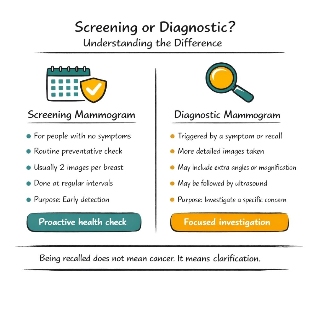
The core difference is intent: screening is a proactive check for those without symptoms, while diagnostic is a reactive, detailed look into a specific concern.
Knowing the difference helps set your expectations. A screening mammogram is a routine part of preventative healthcare, usually recommended for women starting from age 40 or 50, depending on national guidelines. Think of it in the same way as other early-detection tests, like those that measure the prostate-specific antigen for men, which serve a similar purpose for different health conditions.
If you get a call-back after a screening, it simply means something needs a closer look. This next step is usually a diagnostic mammogram, where the technologist will take more pictures, perhaps from different angles or zooming in on one particular spot. The goal is just to gather more information for a clear and accurate picture, helping to provide peace of mind and guide whatever comes next.
Knowing what to do before your appointment can make the whole experience feel much smoother and less daunting. Honestly, a little bit of prep work can make a world of difference, helping you walk in feeling confident and ensuring we get the clearest possible images.
It’s completely normal to feel a bit anxious before a mammogram. Lots of people do. If you're feeling nervous, simple relaxation practices can really help take the edge off. You might want to look into some meditation techniques for beginners to help find a little calm beforehand. Just taking a few quiet moments for yourself can work wonders.
Beyond managing any pre-scan jitters, there are a few practical things to keep in mind on the day.
Here's a tip I always share with my patients: try to schedule your mammogram for the week after your period. Your breasts are usually least tender during this time, which can make the compression part of the exam a lot more comfortable.
As for what to wear, think simple and practical.
Planning these small details helps everything run smoothly once you arrive, letting you focus on the appointment itself.
This one is really important. On the morning of your mammogram, do not apply deodorant, antiperspirant, lotion, cream, or powder anywhere on your chest or under your arms.
Why does this matter so much? Many of these products contain tiny metallic particles, usually aluminium-based. While they're perfectly safe on your skin, they can show up on the X-ray as small white spots. The problem is, these spots can look a lot like calcifications, which can be an early sign of breast cancer. This could lead to an unclear result, a lot of unnecessary worry, and the hassle of coming back for more tests.
By skipping these products for just one day, you help ensure your mammogram images are as clear and accurate as they can be, avoiding any confusion for the radiologist.
The technologist (or mammographer) performing your scan is there to guide you through everything. They’re a trained professional whose job is to get the best quality images while keeping you as comfortable as possible.
Don’t be shy about talking to them. Before you start, make sure you mention:
Giving them this heads-up helps them understand what’s going on with your body and tailor the exam to your needs. It’s all part of making sure you get the best care and the most accurate results possible.
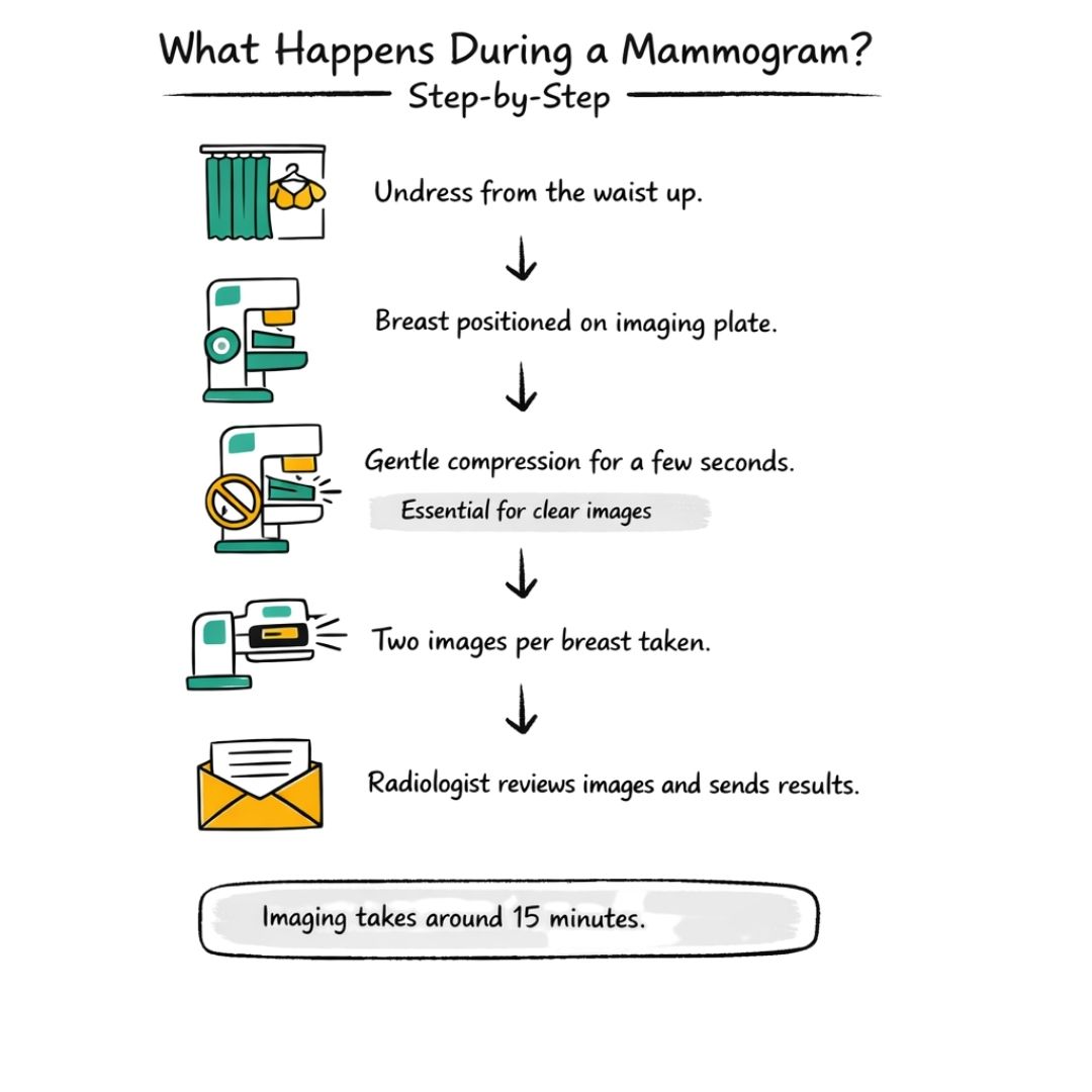
Walking into the mammogram room for the first time can feel a little daunting, but knowing exactly what’s involved can make a world of difference. Let's walk through the process step-by-step, so you feel confident and prepared when you arrive.
You’ll be guided through everything by a specially trained radiographer or technologist. Their job is to make you feel as comfortable as possible while getting the clearest images they can. They are there to help, so don't hesitate to ask them anything.
The whole thing is surprisingly quick. A standard screening mammogram usually takes less than 15 minutes for the imaging itself, with the total appointment lasting about 30 minutes.
First, the technologist will help you stand in front of the mammogram machine. You'll place one breast on a special platform, and then a clear plastic plate will slowly lower to gently but firmly compress your breast for a few seconds.
This compression is probably the most talked-about part of the procedure, and for good reason—it’s absolutely essential for getting a clear picture.
Think of it like this: trying to see through a thick, cloudy glass of water is hard, but if you pour that water into a thin, flat dish, you can see every tiny speck. Compression spreads out the breast tissue in the same way, making sure no small details are hidden.
It also holds the breast perfectly still, which prevents any blurring. While the pressure can be uncomfortable, it only lasts for a few seconds for each image. Most people describe it as a firm squeeze. If you ever feel sharp pain, please tell the technologist right away so they can readjust.
For a routine screening, the technologist typically takes two X-ray images of each breast. One view is taken from top to bottom, and another is taken from the side at an angle. This gives the radiologist a complete picture of all your breast tissue.
You’ll be asked to hold your breath and stay still for the few seconds the X-ray is being taken. The technologist will step behind a screen while the image is captured, but they’ll be right back to help you get into position for the next one.
This simple infographic is a great reminder of what to do on the day.
Following these small steps makes a big difference in ensuring the process is smooth and the results are accurate.
Once all the images are taken, the technologist will do a quick check to ensure they are high quality and not blurry. If needed, they might have to retake one. As soon as they give the all-clear, you’re all done and can get dressed.
The images are then passed to a radiologist, a doctor who specialises in reading medical images like X-rays and scans. They will meticulously examine your mammogram for any unusual signs. This is similar to how other imaging tests work; for instance, you can read about what happens during a pancreas ultrasound in another of our guides.
You and your doctor should receive the results within a couple of weeks, although this can vary. We’ll cover how to interpret these results and what happens next in the following section.
Let’s be honest, waiting for your mammogram results can be the most nerve-wracking part of the whole experience. When that letter or message finally lands, the technical language can feel a bit like reading another language, which doesn't help with the anxiety. Let's walk through what the results really mean, so you feel more informed and in control.
Most of the time, the news is good. A 'negative' or 'normal' result is the all-clear—it means the radiologist didn’t spot anything concerning. You can breathe a sigh of relief and just stick to your regular screening schedule.
Sometimes, though, you might get a call asking you to come back for more tests. This is known as a ‘recall’, and it's understandably a moment that can make your heart sink.
Getting a recall notice is scary, but the first thing to know is that it’s incredibly common and does not mean you have cancer. Far from it. In fact, fewer than 1 in 10 women who are recalled for more tests are actually diagnosed with breast cancer.
A recall simply means the radiologist saw something on the first scan that wasn't perfectly clear and needs a closer look. It's about being thorough.
Here are a few common reasons for a callback:
Think of a recall as the screening process working exactly as it should. It’s a sign of caution and thoroughness, designed to make sure nothing is missed. It's a step for clarification, not a diagnosis.
If you are called back, the follow-up appointment is much more focused. The goal is simply to get a sharper, more detailed picture of the specific area that raised a question.
You will likely have one or more of these tests:
These follow-up tests usually give the medical team all the information they need to confirm that everything is fine. If they do find something that needs a closer look, like a potential case of lobular breast cancer, they will explain the next step, which is usually a biopsy.
It's worth remembering the incredible impact of screening programmes. For example, the NHS Breast Screening Programme in the UK has played a huge part in improving outcomes. It has helped lift breast cancer survival rates from around 40% in the 1970s to about 76% more recently. If you're interested, you can read the full research about breast cancer survival rates to learn more about the difference screening makes.
While a standard mammogram is a powerful screening tool, it's not a one-size-fits-all procedure. Certain factors, like having dense breast tissue or implants, mean your screening might need a slightly different approach.
If this sounds like you, don't worry. It simply means your radiology team will use some specialised techniques to make sure they get the clearest possible picture of your breast health.
Breasts are a mix of supportive tissue (fibrous and glandular) and fatty tissue. Breast density is just a term for how much of that supportive, fibrous tissue you have compared to fatty tissue. On a mammogram, fatty tissue shows up as a transparent dark grey, while dense tissue appears as solid white.
Here’s the challenge: potential cancers also show up as white areas. In very dense breasts, this can create a camouflage effect, making it harder to spot subtle changes. It's like trying to find a polar bear in a snowstorm. This is why women with dense breasts are often offered additional screening tests.
Having dense breasts is incredibly common—it’s not a disease or anything you’ve done wrong. It’s mostly down to genetics and hormones. While it doesn't mean you have cancer, it is considered a small risk factor.
Recent research is making great strides in this area. In the UK, for instance, about 10% of women have extremely dense breasts, and nearly 50% fall into the dense category. A major trial found that using extra imaging techniques could more than triple the cancer detection rate for these women. You can read more about this promising work on enhanced breast cancer screening on cam.ac.uk.
If you’re told you have dense breasts, your doctor might recommend another type of scan to get a better look.
Breast Ultrasound: This uses sound waves to build a picture of the breast tissue. It’s brilliant for telling the difference between a harmless fluid-filled cyst and a solid lump. Learning more about how ultrasound can detect cancer can help you feel more prepared for this step.
Breast MRI: An MRI uses powerful magnets to create incredibly detailed images. It’s usually reserved for women with a very high risk of breast cancer but is also a valuable tool for assessing dense tissue.
Getting a mammogram with breast implants is perfectly safe, but it does require a bit of extra skill. When you book your appointment, make sure you let the clinic know you have implants. They’ll ensure you’re scheduled with a technologist who has plenty of experience with this.
The goal is to get a clear image of the natural breast tissue around the implant. To do this, the technologist uses a special method called the Eklund displacement view. They’ll gently push the implant back towards your chest wall while carefully pulling your breast tissue forward to be imaged.
This allows the X-ray to capture just your natural tissue, without the implant getting in the way. People who have had a mastectomy often explore different breast reconstruction options after mastectomy, and implants are a common choice. Knowing how to monitor them effectively is key. You'll have a few extra images taken, but it’s all to ensure your screening is as thorough as possible.
To give you a quick overview, here’s how mammograms are adapted for dense breasts and implants.
| Consideration | Challenges | Solutions & Techniques |
|---|---|---|
| Dense Breast Tissue | Dense tissue appears white on a mammogram, which can hide or "camouflage" small cancers that also appear white. | Supplemental Screening: Using ultrasound or MRI to get a clearer picture. Tomosynthesis (3D mammography) can also improve detection. |
| Breast Implants | Implants can block the view of some breast tissue, making it difficult to see everything clearly. | Eklund Displacement Views: A specialised technique where the implant is pushed back to allow better imaging of the natural breast tissue in front. |
Ultimately, whether you have dense breasts or implants, the goal is always the same: to get the most accurate screening possible. By working with an experienced team and using the right techniques, we can achieve just that.
Think of a mammogram as a detailed snapshot, capturing a picture of your breast tissue at a single point in time. It's an incredibly powerful tool, but it's only one part of the bigger picture of your breast health. The most effective approach combines these regular checks with your own day-to-day awareness.
Official screening guidelines are there to give us a structured, evidence-based schedule for these snapshots. It's worth knowing that these recommendations can differ depending on where you live.
In the UK, the NHS Breast Screening Programme usually invites women for their first mammogram somewhere between the ages of 50 and 53. After that, you'll be invited back every three years until you're 71. In contrast, organisations in the USA, like the American Cancer Society, often suggest yearly mammograms for women starting from age 40.
So, why the different timings? These schedules are built on mountains of research to figure out when screening does the most good. They’re carefully designed to weigh up the life-saving benefits of catching cancer early against the small risks, like radiation exposure or the anxiety of a false alarm. Sticking to the recommended schedule for your age group gives you the best odds of spotting any problems when they are smallest and easiest to treat.
While screening provides the timetable, breast awareness is your constant companion. After all, nobody knows your body better than you do. This isn't about following a complicated self-exam routine once a month; it's much simpler. It's just about getting to know the normal look and feel of your breasts.
A mammogram is a test you have every few years, but you live with your body every single day. Being 'breast aware' means you are the first line of defence, ready to notice subtle changes between appointments.
Being breast aware just means you know what’s normal for you. That way, you’re much more likely to spot something out of the ordinary, like:
If you do spot any of these changes, the most important thing is to get in touch with your doctor straight away. Please don't put it off or wait for your next scheduled mammogram. When it comes to breast cancer, catching it early makes a world of difference, and your own awareness is a crucial part of that.
Getting people to attend their screening appointments is a major focus in many countries. In the UK, for instance, the NHS invites women aged 50 to 71 every three years, but recent data shows the uptake rate is around 70% – quite a bit below the NHS target of 80%. These numbers really drive home why it's so vital to go when you're invited. If you'd like to understand more, you can discover more insights about UK breast cancer screening rates and the work being done to encourage more people to take part.
Even when you know what to expect, a few questions can still pop into your head. Let's run through some of the most common queries we hear, giving you quick, clear answers to help put your mind at ease.
Honestly, most people find it more awkward or uncomfortable than outright painful. The machine needs to apply firm pressure to get a clear image, but this only lasts for a few seconds for each picture. It’s certainly a strange sensation.
How much discomfort you feel can really depend on your own sensitivity, and even the time of the month. A great tip is to schedule your appointment for the week after your period finishes, as your breasts tend to be least tender then. And always, always tell the radiographer if you're in any real pain.
Mammograms use a tiny, very safe dose of radiation. To put it into perspective, it's about the same amount of natural background radiation you’d be exposed to over a few months just by going about your daily life.
Health experts all agree that the huge benefit of catching cancer early with a mammogram far outweighs the minimal risk from this small amount of radiation. Modern digital machines are also incredibly efficient, designed to use the lowest dose possible while still getting a high-quality picture.
First, take a deep breath. Getting a callback is surprisingly common and it does not automatically mean you have cancer. In fact, in some screening programmes, around 1 in 20 women are called back for more tests after a routine screening, and for most, it turns out to be nothing to worry about.
It could be something simple like a benign (non-cancerous) cyst, an area of dense tissue that needs a closer look, or the first image just wasn't clear enough. Your follow-up appointment will usually involve more specialised mammogram views or perhaps a breast ultrasound to get more detail.
Think of a callback as the screening process doing its job properly. It’s a sign that they're being extra cautious to double-check anything that looks even slightly unusual. It's a step for clarification, not a diagnosis.
Yes, absolutely. Men don't have routine screening mammograms like women do, but if a man discovers a lump or notices other symptoms like nipple discharge or skin changes, a diagnostic mammogram is often the first imaging test performed.
Although breast cancer in men is rare, making up less than 1% of all cases, it does happen. The procedure for men is very similar to the one for women and is a crucial tool for diagnosis. Facing a potential diagnosis is tough for anyone, and getting the right support is key. For practical advice on managing the journey, you can find helpful resources on coping with cancer.
We strongly advise you to talk with a health care professional about specific medical conditions and treatments.
The information on our site is meant to be helpful and educational but is not a substitute for medical advice.
Body image really suffers from breast cancer and access to a good bra is critical for a woman to revive herself. The connection is intricate.
Asking 'does an ultrasound show cancer?' This guide explains what ultrasound can detect, its limitations, and what the next steps are in the diagnostic process.
Facing a stereotactic breast biopsy? Discover what to expect with our empathetic step by step guide for preparation recovery and support throughout.